Anatomical Name Of Lower Back Muscles / Muscles Move And Support The Spine. This picture also contains humerus, olecranon process of ulna, deep to tendon and so on. The lower part of the trapezius ascends and depresses the scapula, while the transverse or middle region of the trapezius is what retracts the. Anatomical name of lower back muscles : To perform clinical clinical orthopedic manual therapy to the lumbar spine. Each lumbar spinal level is numbered from top to bottom—l1 through l5, or l6.
The lower part of the trapezius ascends and depresses the scapula, while the transverse or middle region of the trapezius is what retracts the. They originate from the thoracolumbar fascia, the spinous process of thoracic six through 12, the iliac crest, and your lower three ribs. In turn, the posterior deep muscles are the piriformis, obturator internus, obturator externus, superior gemellus, inferior gemellus, and quadratus femoris. The quadratus lumborum muscles (orange, in the image above) are found in the lower back (also called the lumbar area). This curve, called lordosis, helps to:
Muscles Of The Posterior Leg Attachments Actions Teachmeanatomy from teachmeanatomy.info The lordotic curve your lower back (lumbar spine) is the anatomic region between your lowest rib and the upper part of the buttock. Anatomical name of lower back muscles : This depth, combined with the fact that the psoas originates from the sides of the five lumbar vertebrae, means it plays an important role in back health. The lower part of the trapezius ascends and depresses the scapula, while the transverse or middle region of the trapezius is what retracts the. The muscle then courses up to your shoulder and attaches to your upper arm bone. Human musculature bodybuilding infographic muscular system vector human anatomy back muscle anatomy bicep male muscular anatomy human body anatomy female female anatomy muscle hamstrings muscle. All the extrinsic back muscles are innervated by the ventral (anterior) rami of the cervical spinal nerves , except for the trapezius muscle which receives its supply from the accessory nerve (cn xi) . Intermediate extrinsic muscles of the back:
The posterior superficial muscles are the three gluteal muscles (gluteus maximus, gluteus medius, gluteus minimus), and the tensor fascia latae.
Lumbar spine lower back and superficial muscles the muscles of the lower back help stabilize, rotate, flex, and extend the spinal column, which is a bony tower of 24 vertebrae that gives the body. The muscle then courses up to your shoulder and attaches to your upper arm bone. There are three parts to the trapezius. When it contracts, it makes the foot bend downward, and it also helps to bend the knee. Related posts of muscles of the lower back and hip diagram muscle anatomy lower extremity. The latissimus dorsi originates from the lower part of the back, where it covers a wide area. (2017, elsevier) should be consulted. The vertebral column of the lower back includes the five lumbar vertebrae, the sacrum, and the coccyx. All the extrinsic back muscles are innervated by the ventral (anterior) rami of the cervical spinal nerves , except for the trapezius muscle which receives its supply from the accessory nerve (cn xi) . The upper back is a complex area containing a number of muscles that perform various actions on the scapulae (shoulder blades) and humerus. This curve, called lordosis, helps to: The posterior superficial muscles are the three gluteal muscles (gluteus maximus, gluteus medius, gluteus minimus), and the tensor fascia latae. The flexor muscles are attached to the front of the spine and enable flexing, bending forward, lifting, and arching the lower back.
It is innervated by anterior rami of spinal nerves, reflecting its embryological origin outside the back. Muscles of the lumbar spine. There are three parts to the trapezius. Lumbar spine lower back and superficial muscles the muscles of the lower back help stabilize, rotate, flex, and extend the spinal column, which is a bony tower of 24 vertebrae that gives the body. In turn, the posterior deep muscles are the piriformis, obturator internus, obturator externus, superior gemellus, inferior gemellus, and quadratus femoris.
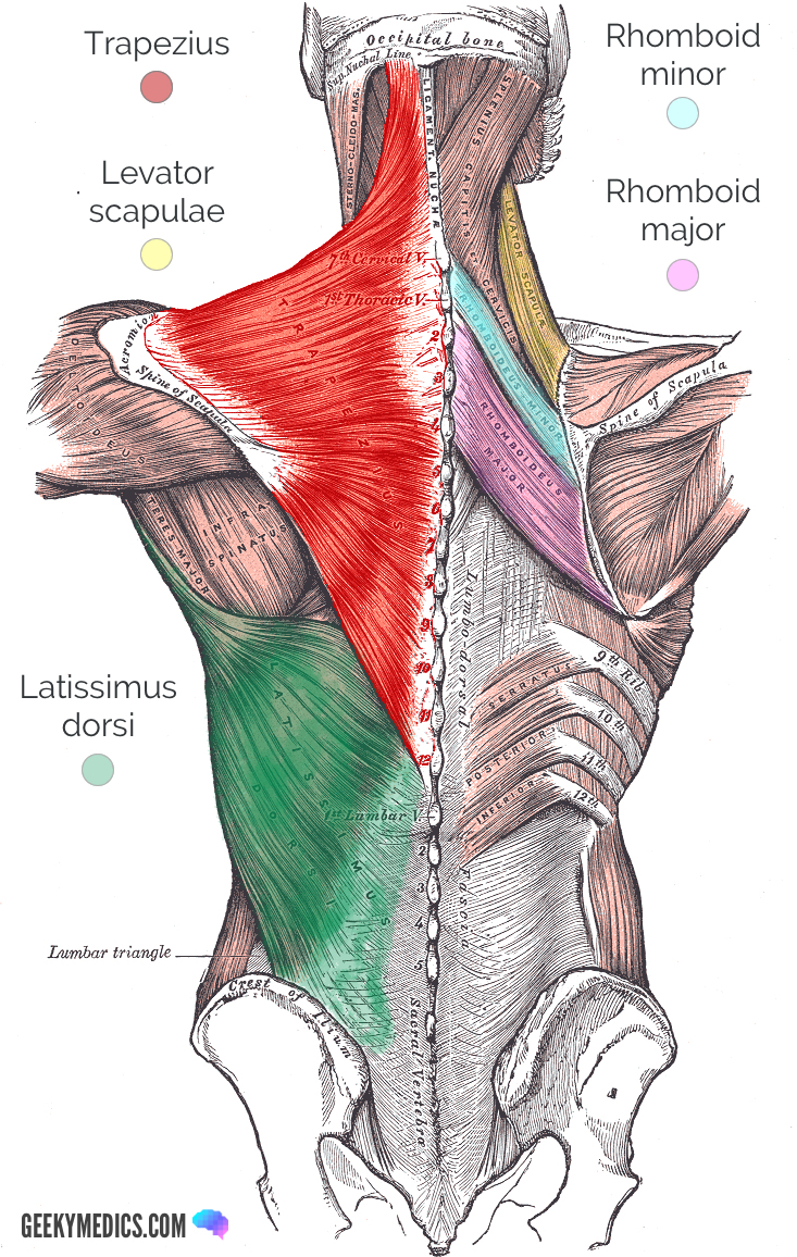
Superficial Back Muscles Anatomy Geeky Medics from geekymedics.com There are three parts to the trapezius. The upper back is a complex area containing a number of muscles that perform various actions on the scapulae (shoulder blades) and humerus. These muscles provide posture and stability to the body by holding the vertebral column erect and adjusting the position of the body to maintain balance. 1 your spine in this region has a natural inward curve. It is composed of trapezius, latissimus dorsi, rhomboid major, rhomboid minor and levator scapulae. On this page, you'll learn about each of these muscles, their locations and functional anatomy. Muscles of the lumbar spine. Related posts of muscles of the lower back and hip diagram muscle anatomy lower extremity.
It is composed of trapezius, latissimus dorsi, rhomboid major, rhomboid minor and levator scapulae.
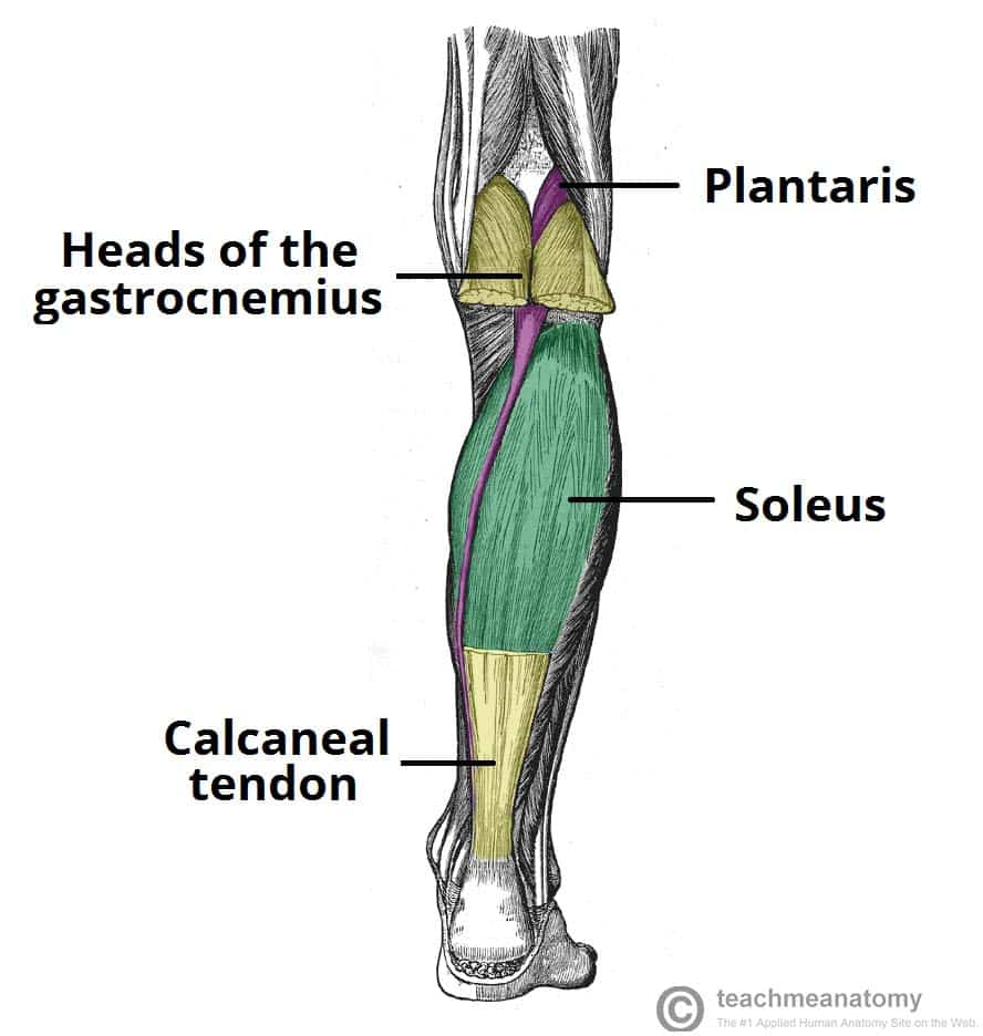
(2017, elsevier) should be consulted. The upper back is a complex area containing a number of muscles that perform various actions on the scapulae (shoulder blades) and humerus. Lumbar spine lower back and superficial muscles the muscles of the lower back help stabilize, rotate, flex, and extend the spinal column, which is a bony tower of 24 vertebrae that gives the body. On this page, you'll learn about each of these muscles, their locations and functional anatomy. These muscles include the large paired muscles in the lower back, called erector spinae, which help hold up the spine, and gluteal muscles. Muscles of the lumbar spine. Its name means belly of the leg,and its common name is the calf muscle. The lordotic curve your lower back (lumbar spine) is the anatomic region between your lowest rib and the upper part of the buttock. The psoas is a hip flexor muscle, as is the quadriceps muscle. Intermediate extrinsic muscles of the back: To perform clinical clinical orthopedic manual therapy to the lumbar spine. As you can see, there are also have a spine of scapula deltoid, triceps brachii, latissimus dorsi. This picture also contains humerus, olecranon process of ulna, deep to tendon and so on.
To perform clinical clinical orthopedic manual therapy to the lumbar spine. This curve, called lordosis, helps to: These muscles provide posture and stability to the body by holding the vertebral column erect and adjusting the position of the body to maintain balance. Your lats are a major back muscle and mover of your shoulder joint. Muscle anatomy chest 12 photos of the muscle anatomy chest anterior chest muscle anatomy, chest muscle anatomy and exercises, chest muscle anatomy male, chest wall muscle anatomy mri, female chest muscle anatomy diagram, human muscles, anterior chest muscle anatomy, chest muscle anatomy and exercises, chest muscle anatomy.
Back Injuries And The Fire Fighter Iaff from www.iaff.org The gastrocnemius runs down the back of the lower leg, from the end of the femur to the heel bone, or calcaneus. There are three parts to the trapezius. The back anatomy includes the latissimus dorsi, trapezius, erector spinae, rhomboid, and the teres major. The superficial muscles of the back are larger and are meant to move the body by producing a lot of force. They help to bend the back to one side or the other. (2017, elsevier) should be consulted. Deep muscles of the back. The psoas muscle is a low back muscle located deep in the body, very close to the spine and inside the hip and thigh bones.
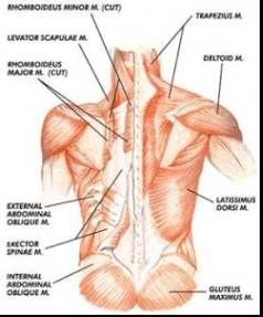
In this image, you will find an occipital bone, sternocleidomastoid, trapezius, deltoid in muscles of the lower back diagram.
The muscles that move the upper legs (thigh) there are many muscles that move the large bone of the thigh. (2017, elsevier) should be consulted. Deep muscles of the back. Related posts of muscles of the lower back and hip diagram muscle anatomy lower extremity. It is composed of trapezius, latissimus dorsi, rhomboid major, rhomboid minor and levator scapulae. The teres majo r muscles work with the rotator cuff muscles to stabilize. Its name means belly of the leg,and its common name is the calf muscle. See back muscle anatomy stock video clips. Serratus posterior superior and serratus posterior inferior muscles. The vertebral column of the lower back includes the five lumbar vertebrae, the sacrum, and the coccyx. Intermediate extrinsic muscles of the back: The trapezius or trapezoid muscles are two paired muscles that extend from the base of the thoracic vertebrae in the spine to the occipital bone and run out to the spine of the scapula. As you can see, there are also have a spine of scapula deltoid, triceps brachii, latissimus dorsi.
Share this post
0 Response to "Anatomical Name Of Lower Back Muscles / Muscles Move And Support The Spine"


0 Response to "Anatomical Name Of Lower Back Muscles / Muscles Move And Support The Spine"
Post a Comment